Some more X-ray pictures

Pictures taken with the DY802 and Kodak Biomax film

I recently tried a film (Kodak Biomax MR) that is used in biotechnology labs for autoradiographs (S-32, P-35), and had good results. Here a sample, a hv solid state rectifier rod. This was exposed for 1 hour at 11cm distance from my best DY802 tube at 10uA/45kV.

I also tried systematicly to find out which exposure time suits best which material. In the follwing sequence of pictures I exposed an iron ring (1mm thick) and a wooden chock (max. height about 1cm) for 5, 10 and 20 minutes. I tried to make the pictures look about as white or black as the real film, and did not artificially increase the contrast.

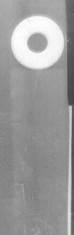
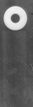
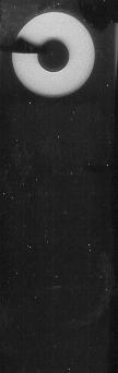
Even after only 5 minutes both ring and chock are both well visible, whereas after 20 minutes the chock is only barely visible. The ring does not change its shade, which means that it absorbs just about all X-ray, whereas the wood absorbs nearly nothing even at its thickest point (towards the ring).

Pictures taken with the PD500 and Agfa Structurix D7 film

Agfa Structurix D7 is a relatively sensitive xray film for non-destructive testing. It was developed at home using standard xray developer (for 10 min.). The following picture was taken at 35cm distance, 10min exposure and 60kV/400uA. It shows a torch and a kitchen alarm clock. The contrast of the original is much better than the scan.

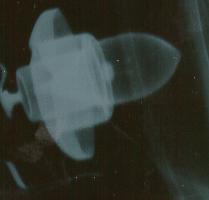
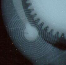
To give you a better impression of the very high resolution of the original, here are two larger scans of details of the alarm clock and torch.

[Back to Index] [Other links]

Jochen Kronjaeger
Kronjaeg@stud-mailer.uni-marburg.de